Magnetic Resonance Cholangio-Pancreatography Scanning Technique
Before we perform scanning, 200-300ml pine apple juice with 1-2ml Gadolinium DTPA (MRI Contrast Media) is given to the patient.Initially a localizer is taken in all three planes with landmark just below xyphoid at rib cage margin. Cover both dome of diaphragm upto the lower level of kidney.
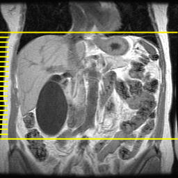
Then, T2 Coronal Single Slice image is planned with breathhold technique and respiratory gating. The epicenter of FOV is placed at center of the image in coronal localizer. The scan line is made parallel to the bile duct or parallel to the cystic duct or perpendicular to the pancreatic duct if possible in axial localizer. Try all of them for better results and in sagittal localizer, the mid point of the vertical scan line is kept at the portal hiatus.

T2 Sagittal Single Slice image is planned now in T2 Coronal Single Slice image with breathhold technique and respiratory gating. The mid point of the slice line is kept at the bile duct and may be angled according to the orientation of the duct.. In axial localizer, the mid point of the vertical scan line is kept at the bile duct (may be less visualized).
T2 Axial STIR sequence is now applied in respiratory gating. Cover from the lower level of dome of diaphragm upto the level of kidney. The mid scan line is kept at the level of cystic duct. or at the level of common bile duct if cystic duct is difficult to access.

T2 Coronal Breathhold Technique sequence is applied then. The mid scan line is kept at the level of bile duct in T2 Axial STIR image. Cover from xiphisternum upto posterior ribs The epicenter of FOV is kept at the level of bile duct.

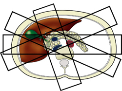
T2 3D TOF sequence is now applied. Set the imaging volume from posterior to the CBD as it passes through the head of the pancreas to anterior to the porta hepatis. Ideally the entire gallbladder should be included but don't worry to exclude the part of gallbladder in order to image the entire CBD. The slab may be angled parallel to the bile duct or perpendicular to the pancreatic duct.


The 3D image is then reconstructed from the raw data obtained.


The routine sequence for Magnetic Resonance Cholangio-Pancreatography are:
T2 Coronal Single Slice Breathhold Technique with respiratory gating
T2 Sagittal Single Slice Breathhold Technique with respiratory gating
T2 Axial STIR with respiratory gating
T2 Coronal with breathhold technique
T2 3D TOF with respiratory gating
Please post your valuable comments!

giving the pineapple juice is an old technique you know..bcos it will reduce the gas shadow but it will induce the secretion of bile and it is not better to scan with an artificial increased bile producing scenario
ReplyDelete