MRI Scanning Technique of Elbow Joint
Initially a localizer is taken in three planes.
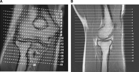
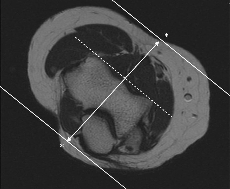
T2 Axial sequence is planned. Use coronal localizer and angle parallel to the elbow joint (parallel to the capitellum and trochlea). Cover from 1 slice distal to radial tuberosity up as far as the slices go (humeral diaphysis).
T1 Axial sequence is planned by copying the slice lines of T2 Axial sequence
 .
.

.
T2 Coronal sequence is planned now. Use axial localizer to angle parallel to the anterior portions of the capitellum and trochlea (or parallel to humeral epicondyles). Use sagittal localizer to angle parallel to humerus/radius/ulnar plane, but closer to plane of radius if minimally flexed (if markedly flexed elbow, then angle between anterior humerus and the radius).

T2 Coronal STIR sequence is planned by copying the slice lines of T2 Coronal sequence.
T2 Sagittal sequence is planned then, where the slice lines are perpendicular to both axial and coronal sequences. Cover 1 slice outside of both humeral epicondyles.
T2 Sagittal STIR sequence is planned by copying slice lines of T2 Sagittal sequence.


The routine MRI sequences for Elbow Joint are:
T2 Axial
T1 Axial
T2 Coronal
T2 Coronal STIR
T2 Sagittal STIR
Please post your valuable comments!

No comments:
Post a Comment