MRI Scanning Technique of Shoulder Joint
Initially a localizer in three plane is taken.


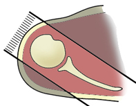
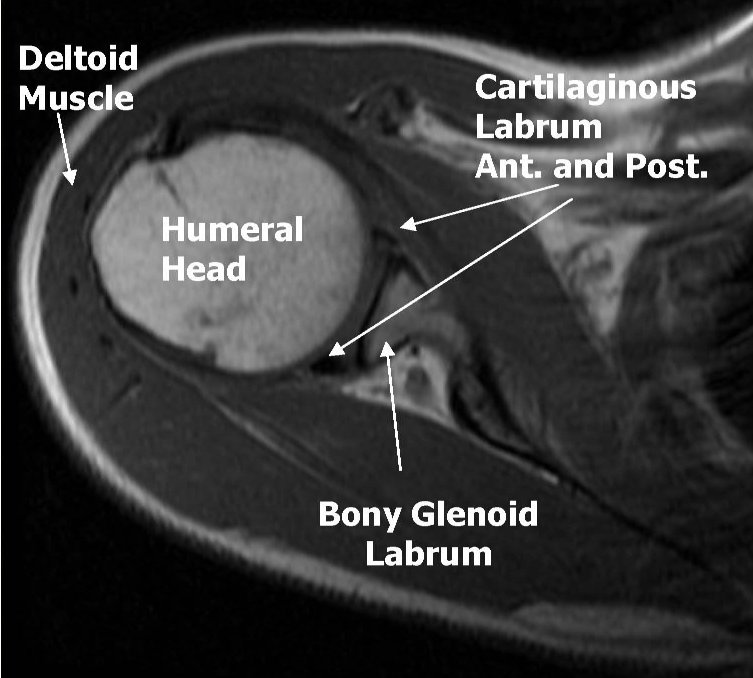
T2 Axial sequence is planned now. Use coronal localizer and the plane is straight horizontal. (If the shoulder is markedly angled, you can angle the axial images perpendicular to the glenohumeral joint). Prescribe plane parallel to humeral shaft. Cover from top of AC joint down and try to cover the inferior portion of glenohumeral joint axillary pouch or the proximal humeral diaphysis.


T1 Axial sequence is planned by copying the slice lines of the T2 Axial sequence.
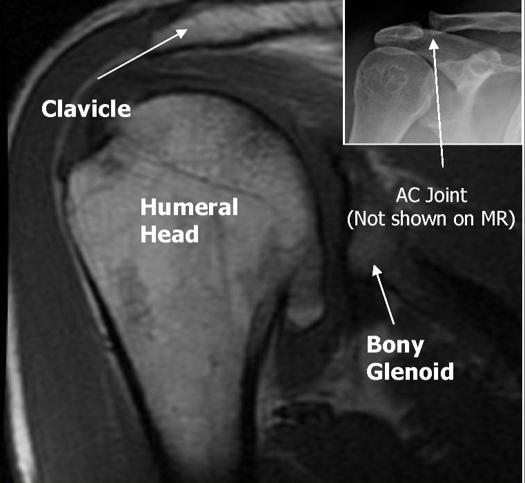
T2 Coronal sequence is planned now. Use Axial image to orient the plane along the supraspinatus tendon. If the supraspinatus tendon is hard to access, we can also use teres minor or the glenohumeral joint. Cover from anterior portion of coracoid process to one slice posterior to the humeral head.


T2 STIR Coronal sequence is planned by copying the slice lines of T2 Coronal sequence.
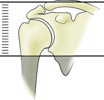
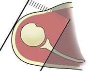
T2 Sagittal sequence is then planned. Angle approximately parallel to the GH joint in T2 Coronal image (Use glenoid articulating surface to angle). Prescribe plane off the axial image with line parallel to bony glenoid. Image from scapular wing to deltoid muscle. Cover from one slice out of humeral head to as far as medial the slices allow (to approximately the medial portion of coracoid process).


ABduction and External Rotation (ABER) results in oblique axial images along the glenoid. The hand is placed behind the head of the supine patient. The superior portion of the glenoid, near the biceps anchor and supraspinatus tendon are well visualized. It also includes the posterosuperior labrum, the midportions of the anterior and posterior labrium and the junction betweent he supraspinatus and infraspinatus tendons, the anterior inferior portion of the glenoid and the infraspinatus and teres minor tendons.


The routine sequence for MRI Scanning of Shoulder Joint are:
T2 Axial
T1 Axial
T2 Coronal
T2 Sagittal
T2 STIR Coronal
Please post your valuable comments!
ha ha perfect copy of my images and protocol...no problem anyway congrats for your efforts to initiate such a great blog...you can always get the protocols and suggestions from me....my email id arun.radiology@gmail.com
ReplyDelete